

MEDICAL VISUAL / PORTFOLIO SAMPLE
Rectal Anatomy: A Visual Guide
An illustrated overview of rectal anatomy, bringing pelvic relationships, wall layers, vessels, nerves and surgical landmarks into one visual.
Surgical trainees, colorectal clinicians and medical educators
Illustrated anatomy guide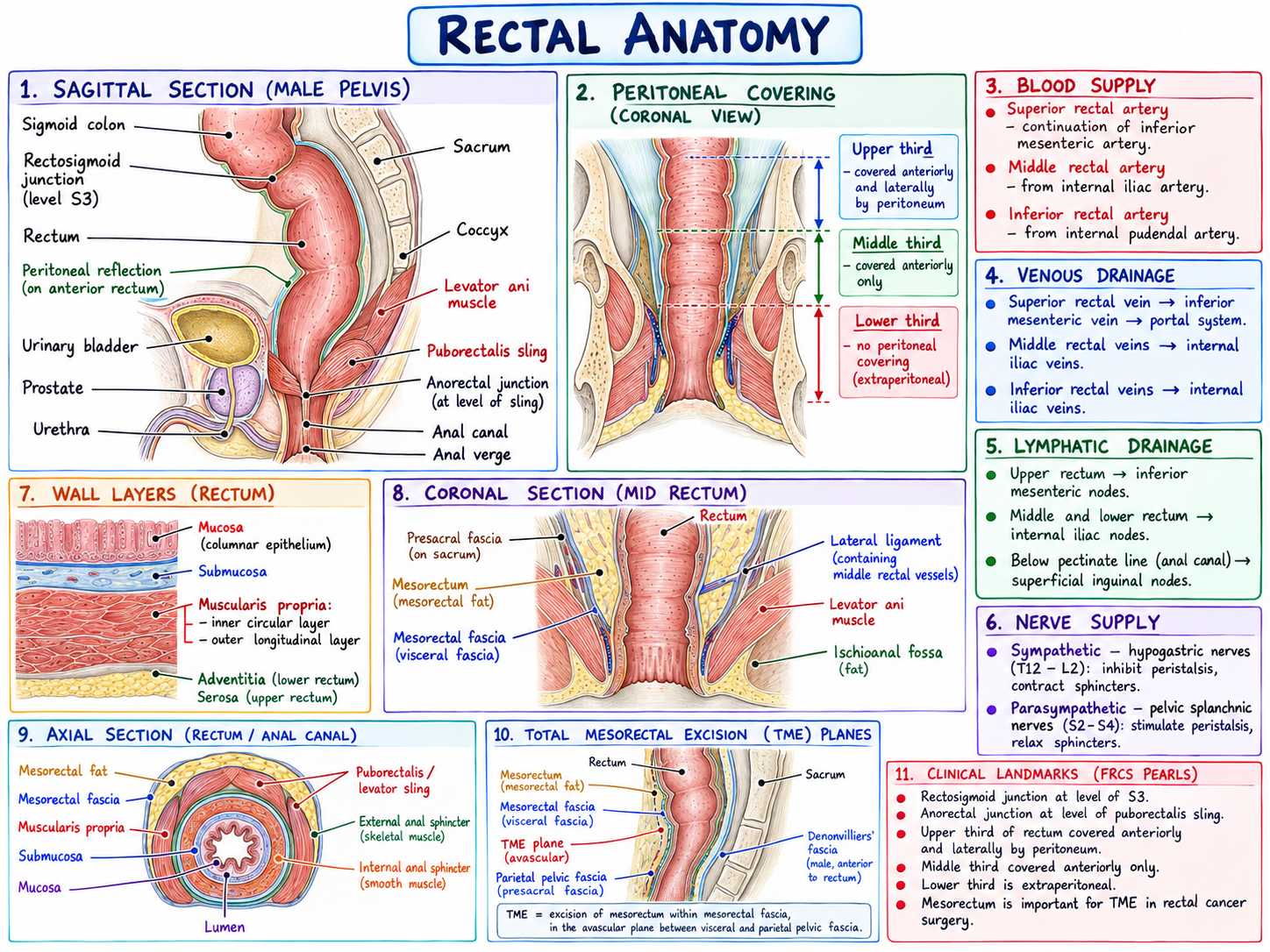
Read the text
RECTAL ANATOMY
Text transcription of the rectal anatomy illustration.
1. Sagittal section (male pelvis)
The labelled structures are the sigmoid colon, rectosigmoid junction (level S3), rectum, peritoneal reflection on the anterior rectum, urinary bladder, prostate, urethra, sacrum, coccyx, levator ani muscle, puborectalis sling, anorectal junction at the level of the sling, anal canal and anal verge.
2. Peritoneal covering (coronal view)
Upper third: covered anteriorly and laterally by peritoneum.
Middle third: covered anteriorly only.
Lower third: no peritoneal covering (extraperitoneal).
3. Blood supply
Superior rectal artery: continuation of the inferior mesenteric artery.
Middle rectal artery: from the internal iliac artery.
Inferior rectal artery: from the internal pudendal artery.
4. Venous drainage
Superior rectal vein to inferior mesenteric vein to portal system.
Middle rectal veins to internal iliac veins.
Inferior rectal veins to internal iliac veins.
5. Lymphatic drainage
Upper rectum to inferior mesenteric nodes.
Middle and lower rectum to internal iliac nodes.
Below the pectinate line (anal canal) to superficial inguinal nodes.
6. Nerve supply
Sympathetic: hypogastric nerves (T12–L2), inhibit peristalsis and contract sphincters.
Parasympathetic: pelvic splanchnic nerves (S2–S4), stimulate peristalsis and relax sphincters.
7. Wall layers (rectum)
Mucosa (columnar epithelium), submucosa, muscularis propria (inner circular and outer longitudinal layers), adventitia (lower rectum) and serosa (upper rectum).
8. Coronal section (mid rectum)
Labels identify the rectum, presacral fascia, mesorectum (mesorectal fat), mesorectal fascia, lateral ligament containing middle rectal vessels, levator ani muscle and ischioanal fossa fat.
9. Axial section (rectum / anal canal)
Labels identify mesorectal fat, mesorectal fascia, muscularis propria, submucosa, mucosa, lumen, puborectalis / levator sling, external anal sphincter (skeletal muscle) and internal anal sphincter (smooth muscle).
10. Total mesorectal excision (TME) planes
Labels identify the rectum, mesorectum, mesorectal fascia, TME plane, parietal pelvic fascia (presacral fascia), sacrum and Denonvilliers’ fascia (male, anterior to the rectum).
The caption describes TME as excision of the mesorectum within the mesorectal fascia, in the avascular plane between visceral and parietal pelvic fascia.
11. Clinical landmarks (FRCS pearls)
Rectosigmoid junction at the level of S3.
Anorectal junction at the level of the puborectalis sling.
Upper third of the rectum covered anteriorly and laterally by peritoneum; middle third covered anteriorly only; lower third extraperitoneal.
Mesorectum is important in TME for rectal cancer surgery.
Portfolio demonstration created by Scalpel & Script. Clinical content should be independently reviewed before use in patient care.
BEHIND THE SAMPLE
From information
to understanding.
This illustrated guide brings several views of the rectum together on one teaching sheet. Sagittal, coronal and axial panels sit alongside concise notes on peritoneal covering, blood supply, drainage and innervation. Wall layers and total mesorectal excision planes give the sample a surgical focus. Handwritten headings and coloured annotations organise the material for revision and teaching. Open the enlarged view to explore the supplied artwork in detail.
- Teaching visual design
- Anatomical illustration
- Information hierarchy
- Revision resource design